
By Dr. Maneesh Dhupper – Gobind Eye Care
Retinal detachment is a vision-threatening eye condition that requires immediate medical attention. If left untreated, it can lead to partial or complete permanent vision loss. Early recognition of warning signs and timely treatment can dramatically improve the outcome.
This guide provides a complete overview of retinal detachment—what it is, its causes, symptoms, diagnosis, and the latest treatment options—to help you protect your vision.
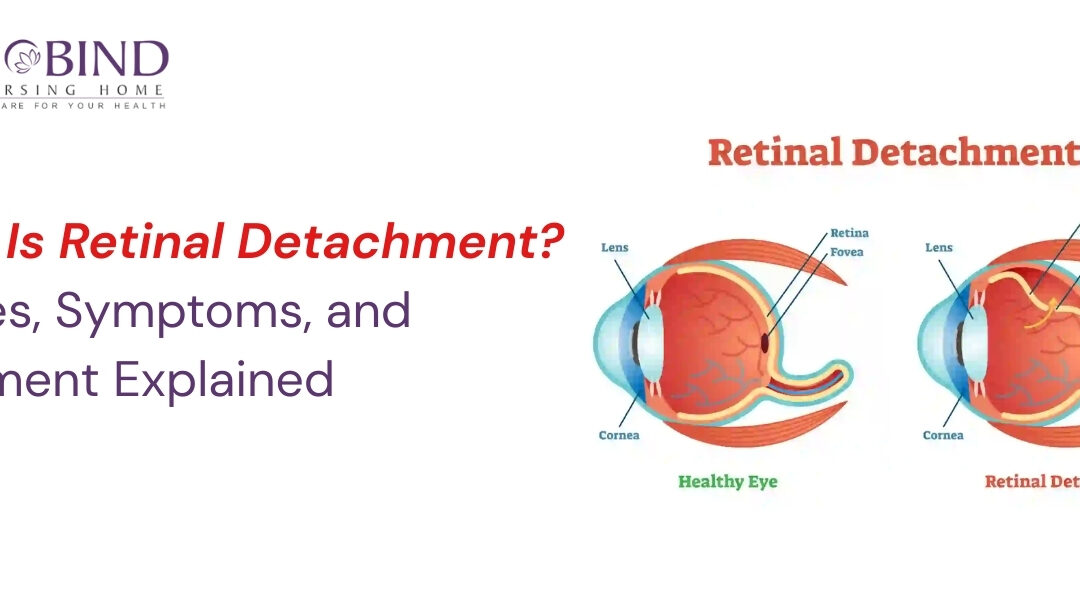
What Is Retinal Detachment?
The retina is a delicate, light-sensitive tissue lining the back of the eye. It functions much like the sensor of a camera—capturing images and sending them to the brain.
Retinal detachment occurs when the retina separates from the underlying supportive tissue.
This separation cuts off the retina’s oxygen and nutrient supply, causing rapid and severe vision impairment.
It is a medical emergency and needs quick intervention to save sight.
Types of Retinal Detachment
1. Rhegmatogenous Retinal Detachment (Most Common)
Caused by a tear, hole, or break in the retina. Fluid seeps through the opening and lifts the retina from its natural position.
Common in older adults, people with high myopia, or those who’ve had eye trauma.
2. Tractional Retinal Detachment
Occurs when scar tissue on the retina contracts and pulls it away from the eye wall.
Frequently seen in advanced diabetic eye disease.
3. Exudative Retinal Detachment
Happens due to fluid buildup under the retina without any tear.
May result from inflammation, tumors, or vascular abnormalities.
Causes and Risk Factors
Retinal detachment can affect anyone, but certain factors increase the risk:
✔ Aging
Natural changes in the vitreous gel can lead to retinal tears.
✔ High Myopia (Nearsightedness)
Longer eye shape and thinner retina increase vulnerability.
✔ Eye Injuries or Trauma
Sports injuries, accidents, or physical impact can create retinal breaks.
✔ Diabetic Retinopathy
Abnormal blood vessels and scarring can cause traction.
✔ Previous Eye Surgery
Cataract surgery or other procedures may slightly raise risk.
✔ Family History
Genetic predisposition can play a role.
Early Warning Symptoms of Retinal Detachment
Retinal detachment is painless, so symptoms must be taken seriously.
Seek immediate help if you notice:
1. Sudden Floaters
Black spots, threads, or cobweb-like shapes drifting in your vision.
2. Flashes of Light
Brief, lightning-like flashes, often at the edges of vision.
3. Blurred or Distorted Vision
Straight lines may look wavy or images appear shadowed.
4. Shadow or Curtain Effect
A dark shade spreading from any side—like a curtain coming down.
5. Sudden Drop in Vision
Sign of advanced detachment requiring urgent treatment.
Early detection is crucial for vision preservation.
How Retinal Detachment Is Diagnosed
An ophthalmologist uses advanced tools to examine and diagnose:
- Dilated retinal examination
- Optical Coherence Tomography (OCT)
- Ultrasound imaging, especially if the view of the retina is blocked
Timely diagnosis significantly enhances treatment success.
Treatment Options for Retinal Detachment
Treatment goals are to reattach the retina and seal retinal breaks. Options include:
1. Laser Photocoagulation
A focused laser creates a seal around retinal tears to prevent detachment.
2. Cryopexy (Freezing Therapy)
Freezing the retinal tear helps it reattach by forming scar tissue.
3. Pneumatic Retinopexy
A gas bubble is injected into the eye to push the retina back into place.
Often used with laser or cryotherapy.
4. Scleral Buckling
A silicone band is placed around the eye to reduce vitreous traction and help the retina reattach.
Useful for complex or large detachments.
5. Vitrectomy
Removes the vitreous gel pulling on the retina and replaces it with gas or silicone oil.
Often used for diabetic or advanced cases.
Can Retinal Detachment Be Prevented?
While not always preventable, risk can be reduced by:
- Regular eye checkups, especially after age 40
- Effective diabetes management
- Protecting eyes from sports injuries
- Paying attention to floaters, flashes, and sudden vision changes
- Monitoring retinal health if you have high myopia
Awareness and early intervention are key.
When to Visit an Eye Specialist
Visit an ophthalmologist immediately if you experience:
- sudden new floaters
- flashes of light
- a curtain-like shadow
- sudden blurred or partial vision loss
Ignoring symptoms may lead to irreversible vision damage.
Conclusion
Retinal detachment is a serious yet treatable condition—if addressed on time. Understanding its signs, causes, and treatment options can help safeguard your eyesight.
If you suspect any symptoms or have high-risk factors, consult an eye specialist without delay.
For expert retinal evaluation and comprehensive eye care services, visit:Dr. Maneesh Dhupper
Also Read:- When Is the Right Time for Cataract Surgery?